
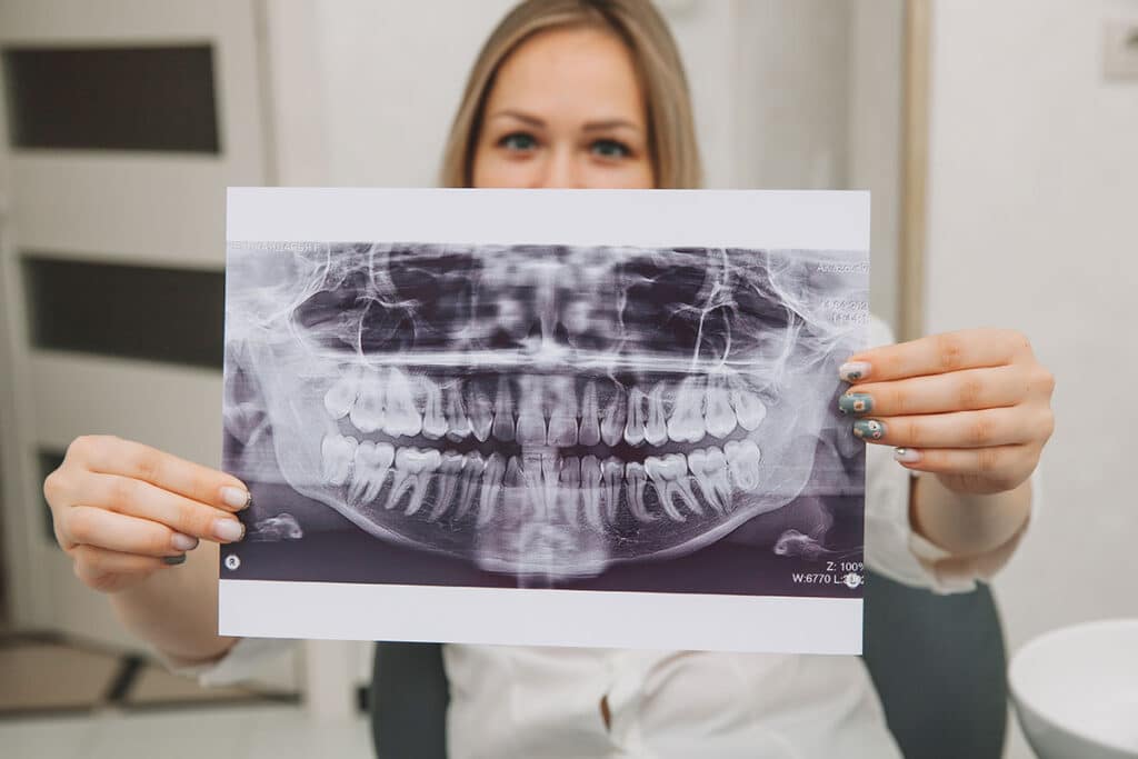
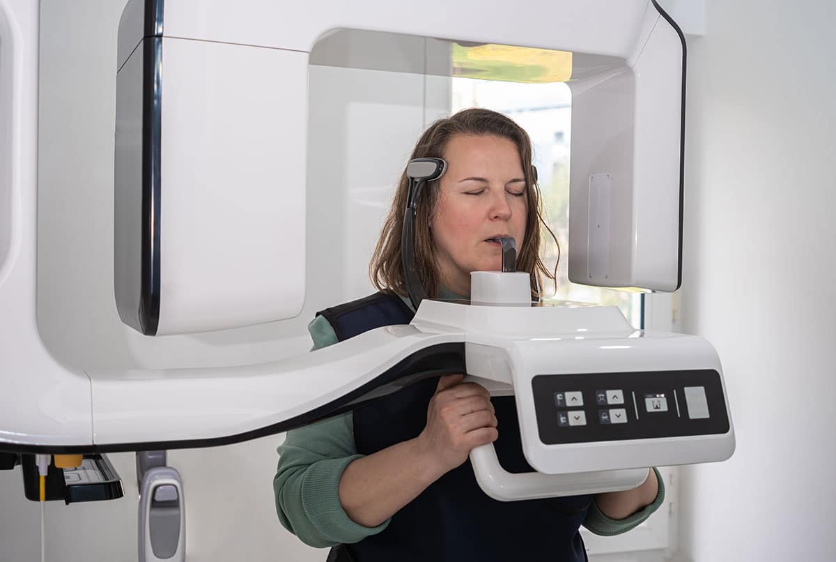
What is a dental X-ray?
Dental X-rays, also known as dental radiographs, are diagnostic images of the teeth, jawbones, and surrounding oral structures that use X-ray technology. Dental X-rays work by using a controlled source of X-ray radiation to create images that your dentist can use to inspect the hidden parts of your mouth.
The best way to explain how a dental X-ray works is to go through the process step by step:
1. X-ray Machine
The dentist or dental radiographer uses a specialized X-ray machine designed for dental imaging. This machine produces a focused beam of X-ray radiation.
2. X-ray Sensor or Film
To capture the X-ray image, a sensor or film is placed in the mouth near the area being examined. In traditional film-based X-rays, a small piece of X-ray film is positioned inside the mouth. In digital radiography, a digital sensor is used, which is similar in size and shape to traditional film but captures digital images.
3. X-ray Exposure
The X-ray machine is positioned outside the mouth, and the X-ray beam is directed at the area of interest. The X-rays pass through the oral tissues and strike the sensor or film. The dense tissues absorb some X-rays, while others pass through and create an image on the sensor or film.
4. Image Formation
In traditional film-based X-rays, the exposed film is then developed in a chemical solution to create a visible image. In digital radiography, the X-ray sensor instantly converts the X-ray energy into a digital image that can be viewed on a computer monitor.
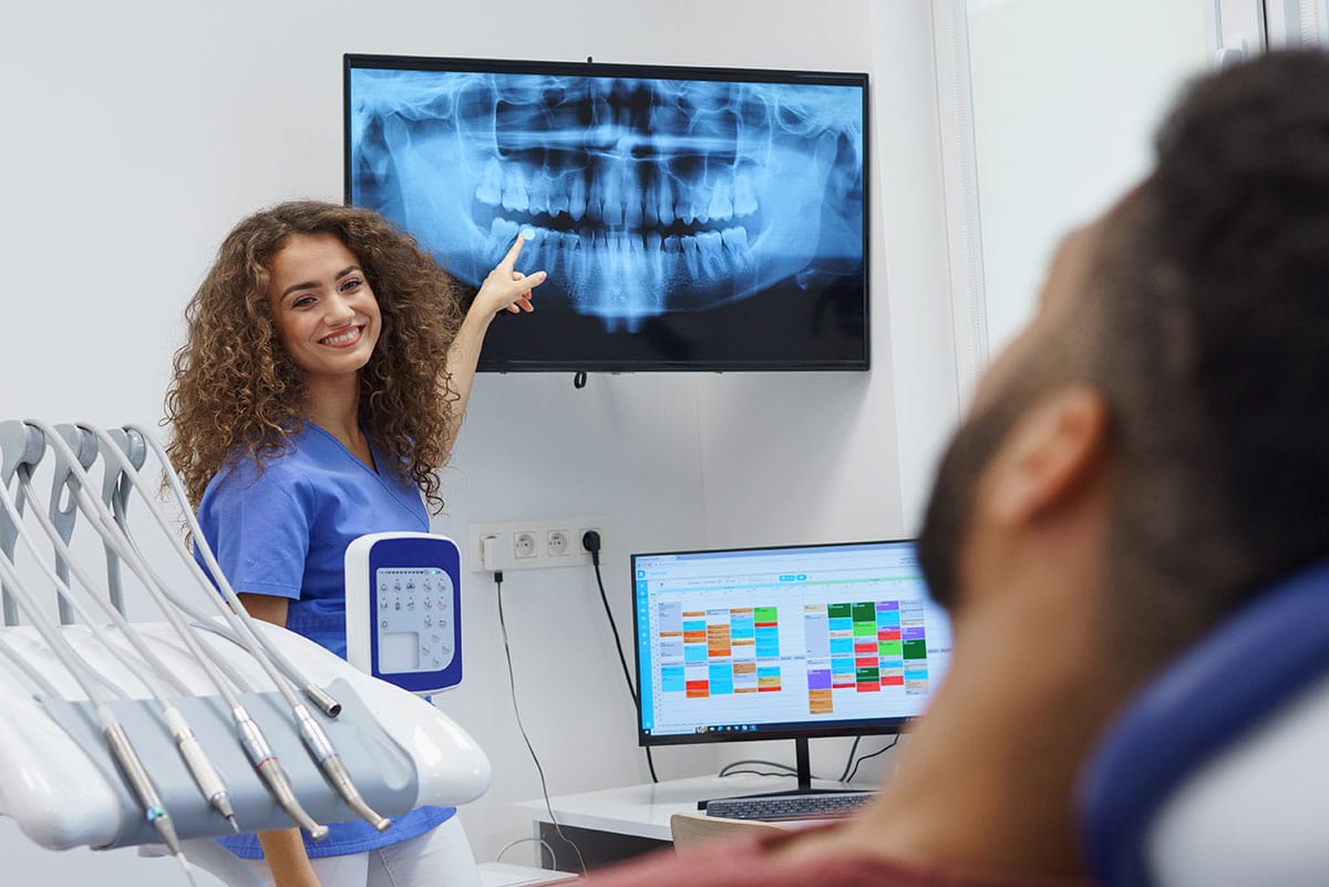
5. Image Viewing
The dentist or dental radiographer views the resulting X-ray images on a computer screen. They can be manipulated digitally to enhance clarity and provide a detailed view of the teeth and oral structures.
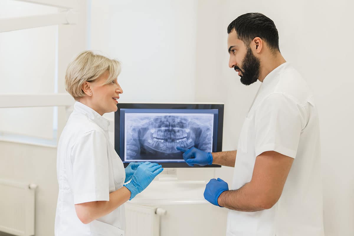
6. Diagnosis and Treatment Planning
The dentist examines the X-ray images to assess the condition of the teeth, roots, bone, and other structures. This helps diagnose oral health issues, plan treatments, and monitor changes over time.
Are there alternatives to dental X-rays?
Dental X-rays are a valuable diagnostic tool in dentistry, but there are alternatives and supplementary methods that may be used depending on the specific situation and the information required:
Visual Examination
Dentists routinely perform visual examinations of the teeth and oral tissues. They can often detect cavities, gum disease, oral lesions, and other issues through careful inspection. However, not all dental problems are visible to the naked eye.
Digital Impressions
Digital impressions are used to create highly detailed 3D models of the teeth and oral structures. They are commonly used in orthodontics and for creating dental prosthetics like crowns and bridges.
These small, handheld cameras can capture high-resolution images of the inside of the mouth. They are often used for patient education and to document the condition of the teeth and gums.
Transillumination
A special light is used to illuminate teeth, allowing dentists to detect cavities and cracks in the enamel. Transillumination is particularly useful for diagnosing early-stage cavities.
Laser Technology
Dental lasers can be used for various purposes, including cavity detection, soft tissue treatments, and the removal of tooth decay. Some lasers can provide valuable diagnostic information.
Saliva Testing
Saliva tests can be used to assess the presence of specific bacteria associated with oral health issues. While not a direct alternative to X-rays, it can provide information about the risk of developing certain conditions.
MRI and CT Scans
In some cases, medical imaging methods like magnetic resonance imaging (MRI) or computed tomography (CT) scans may be used to assess oral and maxillofacial structures. These are typically reserved for more complex cases.
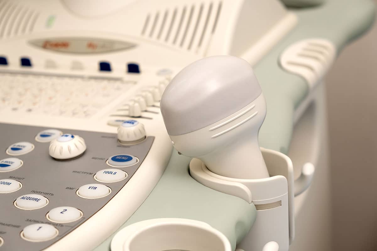
Ultrasound
Ultrasound imaging is occasionally used for dental and maxillofacial purposes, especially in pediatric dentistry or when X-rays are contraindicated.
These alternatives can be useful in specific situations. Still, dental X-rays remain the main tool for diagnosing many dental conditions, especially those involving tooth roots, bone, and areas not visible to the naked eye. The choice of diagnostic method depends on the individual patient’s needs and the dentist’s clinical judgment.
What is digital imaging?
Digital imaging refers to capturing, storing, and displaying visual information in digital format. It has revolutionized various fields, including photography, medicine, dentistry, and many others, by replacing traditional analog techniques with electronic technology. Here’s how digital imaging works and its key advantages:
How digital imaging Works:
1. Image Capture
In digital imaging, an electronic sensor or detector captures visual information, such as photographs, X-rays, or other images. The sensor converts light or radiation (e.g., X-rays) into electrical signals.
2. Analog-to-Digital Conversion
The electrical signals generated by the sensor are then digitized using an analog-to-digital converter (ADC). This process involves converting the continuous analog signal into discrete digital data that a computer can store and process.
3. Digital Storage
The digitized image data is stored on electronic media, such as computer hard drives, solid-state drives, memory cards, or cloud storage. This digital data can be easily duplicated and preserved without degradation in quality over time.
4. Image Processing
Digital images can undergo various processing steps using software tools. This includes enhancing image quality, adjusting colours, removing imperfections, and more. Image processing allows for greater flexibility and control over the final result.
5. Display and Distribution
Digital images can be displayed on electronic screens, such as computer monitors, tablets, smartphones, or specialized medical or dental displays. They can also be easily shared and distributed via email, web pages, and other digital platforms.
Advantages of Digital Imaging
Immediate Results
Digital imaging provides instant access to images, eliminating the need for time-consuming film processing.
Enhanced Image Quality
Digital images can be manipulated to improve clarity and detail, making them valuable for diagnosis and analysis.
Easy Storage and Retrieval
Digital images can be stored electronically, allowing for efficient organization and quick retrieval.
Remote Access
Digital images can be accessed remotely, enabling telemedicine and remote consultations.
Reduced Environmental Impact
Digital imaging eliminates the need for chemicals and film associated with traditional analog methods, reducing environmental waste.
Lower Costs
Over time, digital imaging can be cost-effective, as it reduces the need for film, chemicals, and physical storage space.
Digital imaging has significantly advanced fields such as medical and dental diagnostics, photography, astronomy, and more. It has made it possible to capture, store, and share visual information with unprecedented convenience and precision.
Why are dental X-rays and digital imaging essential?
Dental X-rays are an indispensable tool in modern dentistry, pivotal in maintaining and improving oral health. These diagnostic images provide dentists with invaluable insights into the hidden aspects of oral structures, enabling them to make informed diagnoses and treatment decisions.
Dental X-rays help dentists detect oral health problems early on when they are easier to treat and enable a comprehensive examination. They are an indispensable tool when assessing wisdom teeth concerns, orthodontic treatments, periodontal disease, and treatments like dental implants. Dental X-rays and digital imaging are also often used as visual tools to educate patients about what’s going on with their oral health.
Are dental X-rays safe?
Dental X-rays are considered safe when used with appropriate precautions and modern equipment. The benefits of dental X-rays in diagnosing and monitoring oral health conditions generally outweigh the minimal risks associated with radiation exposure.
Modern equipment produces a very low radiation exposure, and it does it so quickly that there is almost cause for concern, especially when used sparingly and with appropriate safety precautions like lead shields in place.
Even though dental X-rays are usually the most effective and easiest option, if you don’t feel confident about X-rays, most dentists can offer an alternative form of imaging if applicable.
Are dental X-rays safe during pregnancy?
There is always an added level of concern for any dental treatment during pregnancy; dental imaging is no exception. Dental X-rays are generally safe during pregnancy when all necessary precautions (lead apron and collar, limited exposure time) are taken to minimize radiation exposure to the developing fetus. However, if possible, treatment timing of elective dental x-rays should be scheduled before or after the pregnancy. If a dental emergency requires X-rays, you don’t need to worry; they are quite safe for you and your unborn child.
Are dental X-rays safe for children?
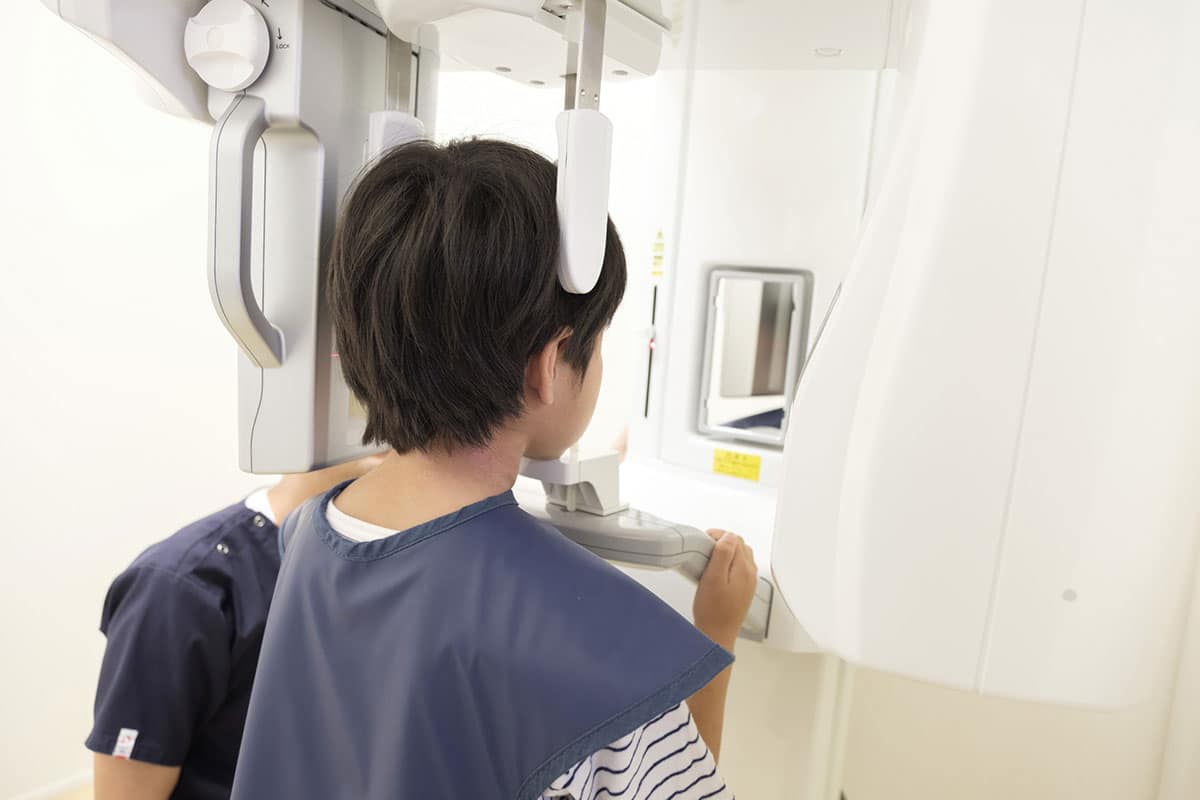
As for pregnant mothers, dental X-rays are also safe for young children, provided all safety precautions are accounted for. Your dentist will, however, still limit the exposure your child receives, but more dental X-rays are generally required for children due to the growing nature of their jaw and teeth – think orthodontic treatment planning.
Should I get a dental X-ray at every appointment?
Whether you should get a dental X-ray at every dental appointment depends on several factors, including your oral health history, risk factors, and the dentist’s recommendation. Dental X-ray frequency is typically determined individually, and no one-size-fits-all approach exists.
Your oral health history, age, current symptoms and other risk factors will determine how often your dentist recommends a dental x-ray.
In conclusion
X-rays, in general, get a bad rap most of the time, which makes sense, seeing as we are dealing with ionizing radiation, even in minuscule doses. There are no wrong questions, and your concerns are completely valid.
That said, modern dental X-rays are completely safe, and the risks associated with improper diagnosing or treatment planning are far greater. While X-rays are still the best way to inspect the hidden parts of your mouth, your dental professional should be more than happy to offer alternatives to dental X-rays if the situation allows.
