
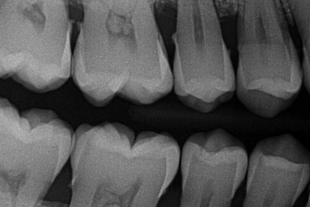
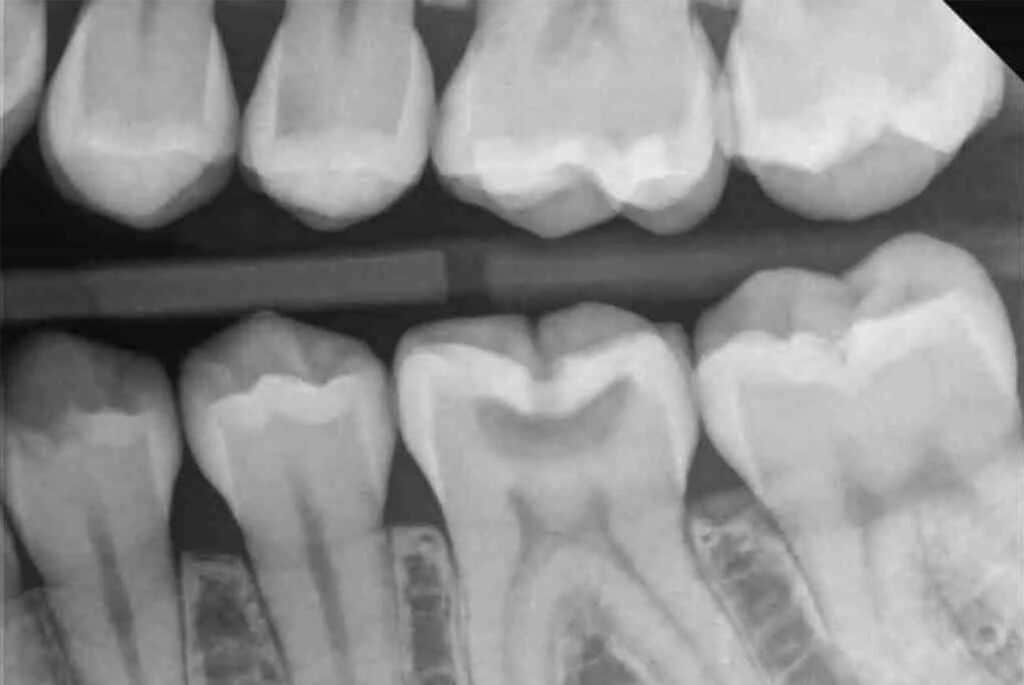


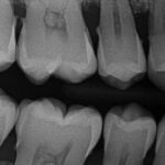
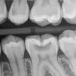
 Probably the most common X-ray we use, these show the upper and lower back teeth in a single view. They are used to detect decay between teeth and changes in bone density caused by gum disease. They are also useful for determining the proper fit of a crown or other restorative device.
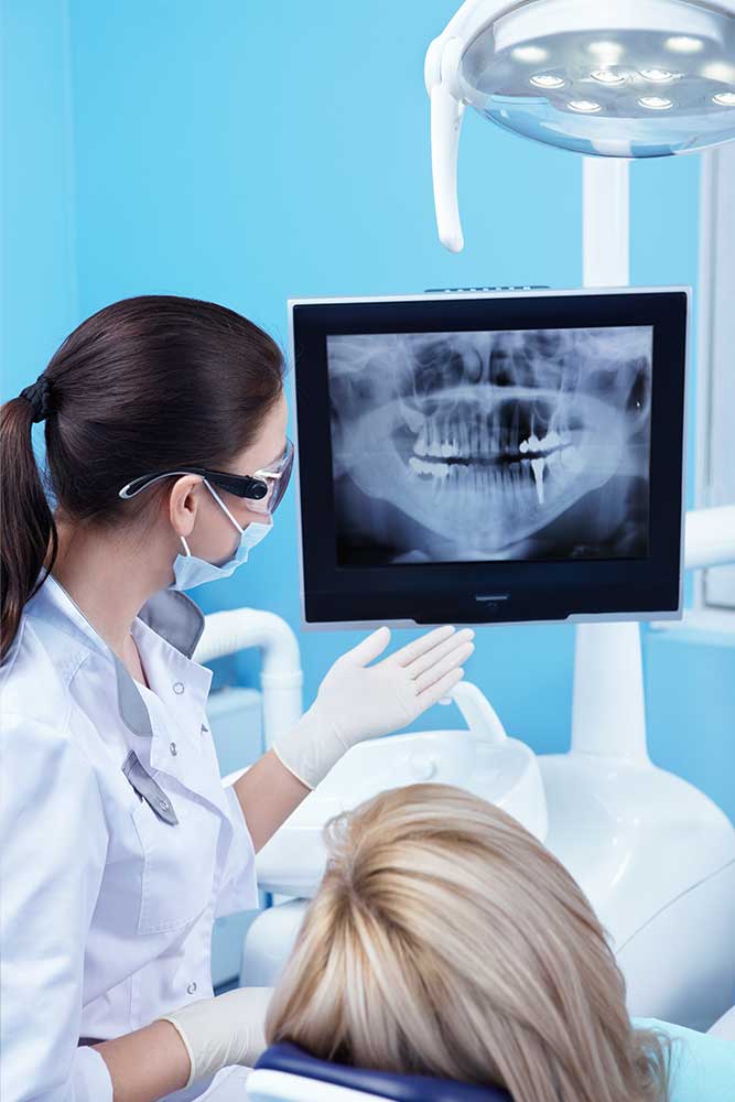
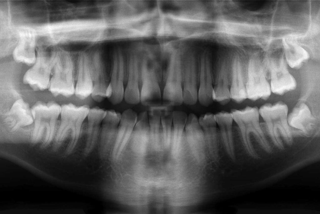
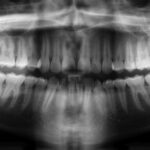 These capture an image of the entire mouth area, showing all the teeth in both upper and lower jaws in a single X-ray. They are useful for detecting impacted teeth, cysts, tumors, jaw disorders, and bone irregularities.
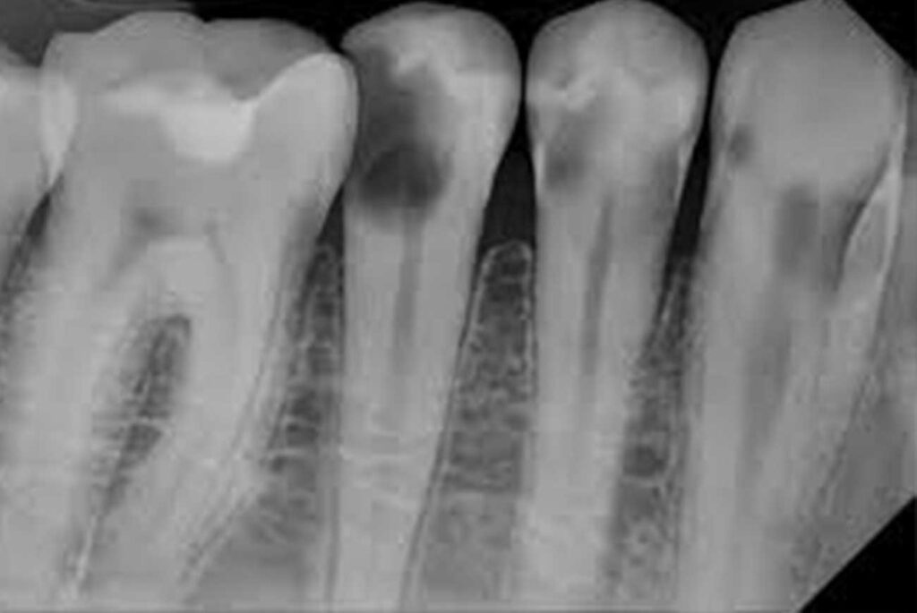
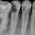 These provide a view of the entire tooth, from the crown to beyond the root where the tooth attaches into the jaw. Periapical X-rays are used to detect dental problems below the gum line or in the jaw, such as impacted teeth, abscesses, cysts, tumors, and bone changes linked to some diseases.
 They are larger and show full tooth development and placement in the upper or lower jaw. These are used to detect the presence of extra teeth, teeth that have not yet broken through the gums, jaw fractures, cleft palate, cysts, abnormalities, or growths.
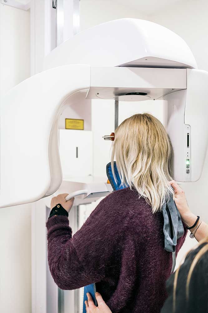
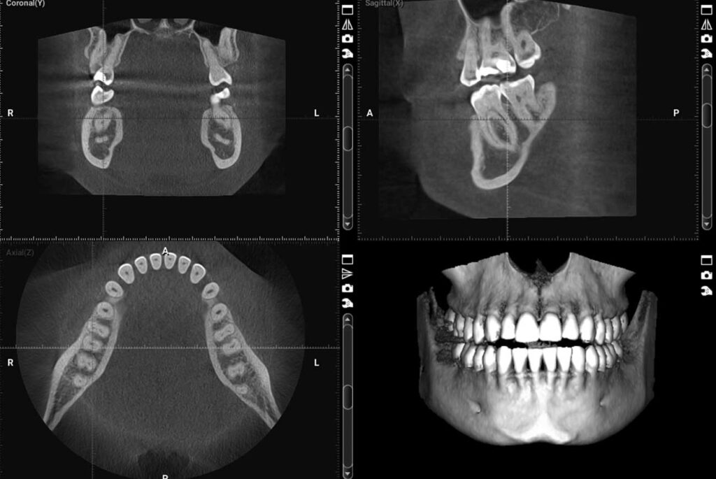
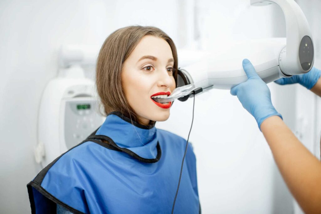
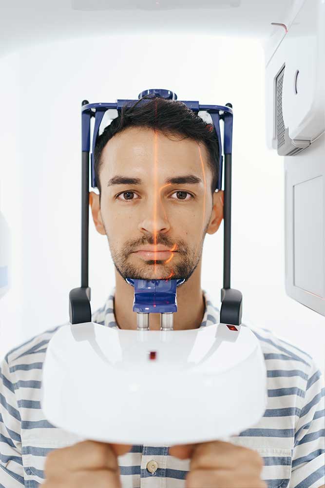
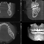 Our most advanced dental X-ray, this technology produces 3D images of the teeth, soft tissues, nerve pathways, and bone in a single scan. It’s used for more complex cases such as implant planning, evaluation of the jaw, sinuses, nerve canals, and nasal cavity, and for detecting and treating orthodontic issues.
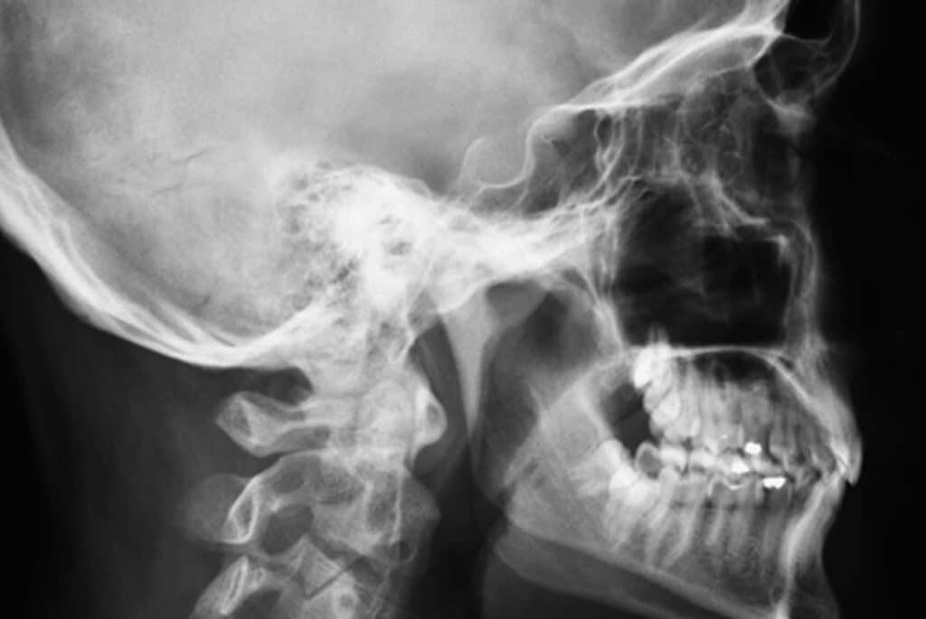
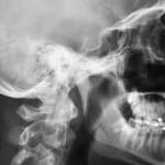 These are commonly used by our orthodontists when planning a treatment schedule because they provide a side view of the face, showing the teeth in relation to the jaw and profile of the individual.
